Bioengineering
October 2024
Modern medicine has made our lives safer
Bioengineering is a fast growing industry. Yet, many challenges still remain to be solved. The success of organ transplantation relies on the availability of scarce donor organs and life-long medication to suppress rejection of the transplant by the patient’s immune system, resulting in a considerable loss of quality of life or even failure of the transplantation.
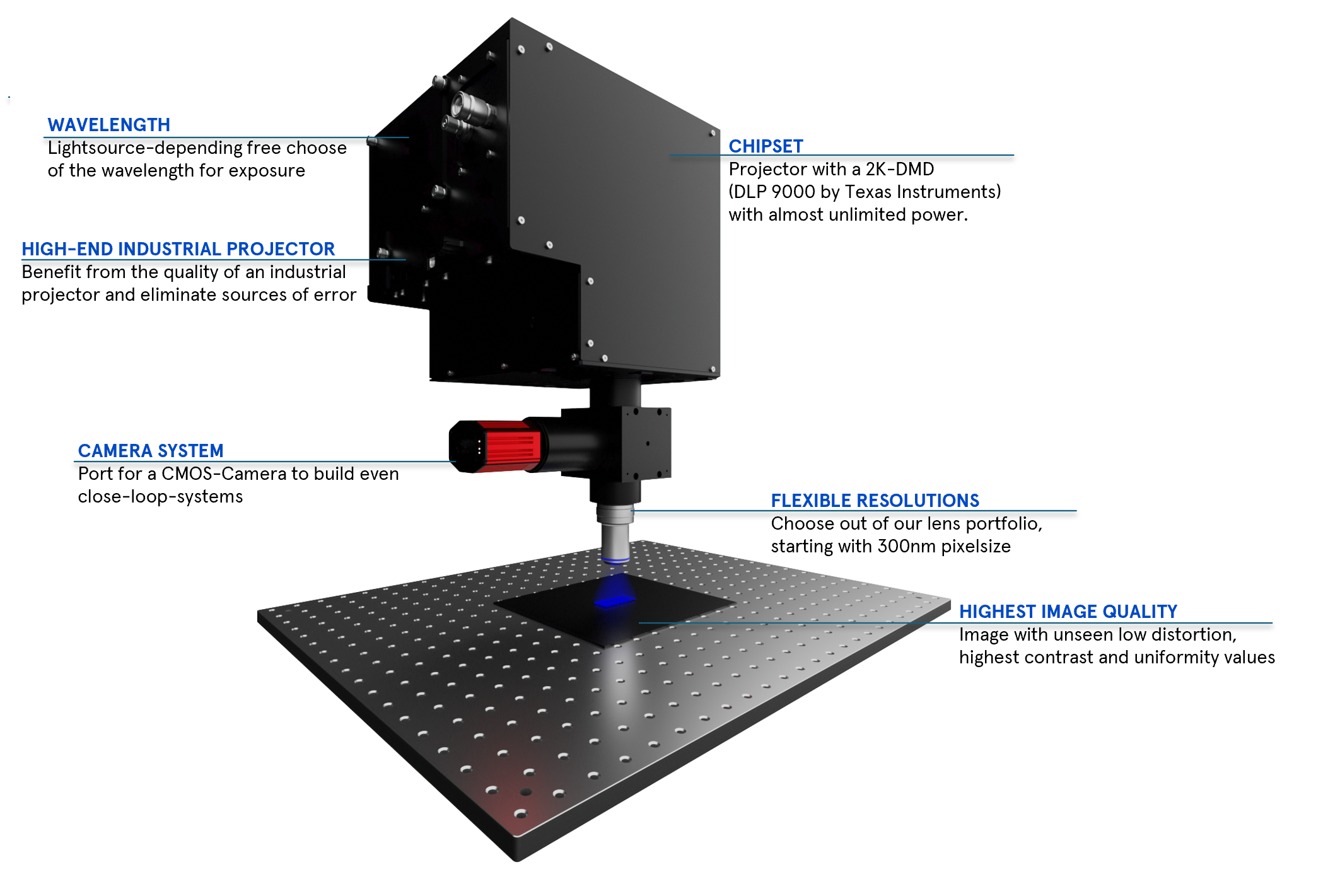
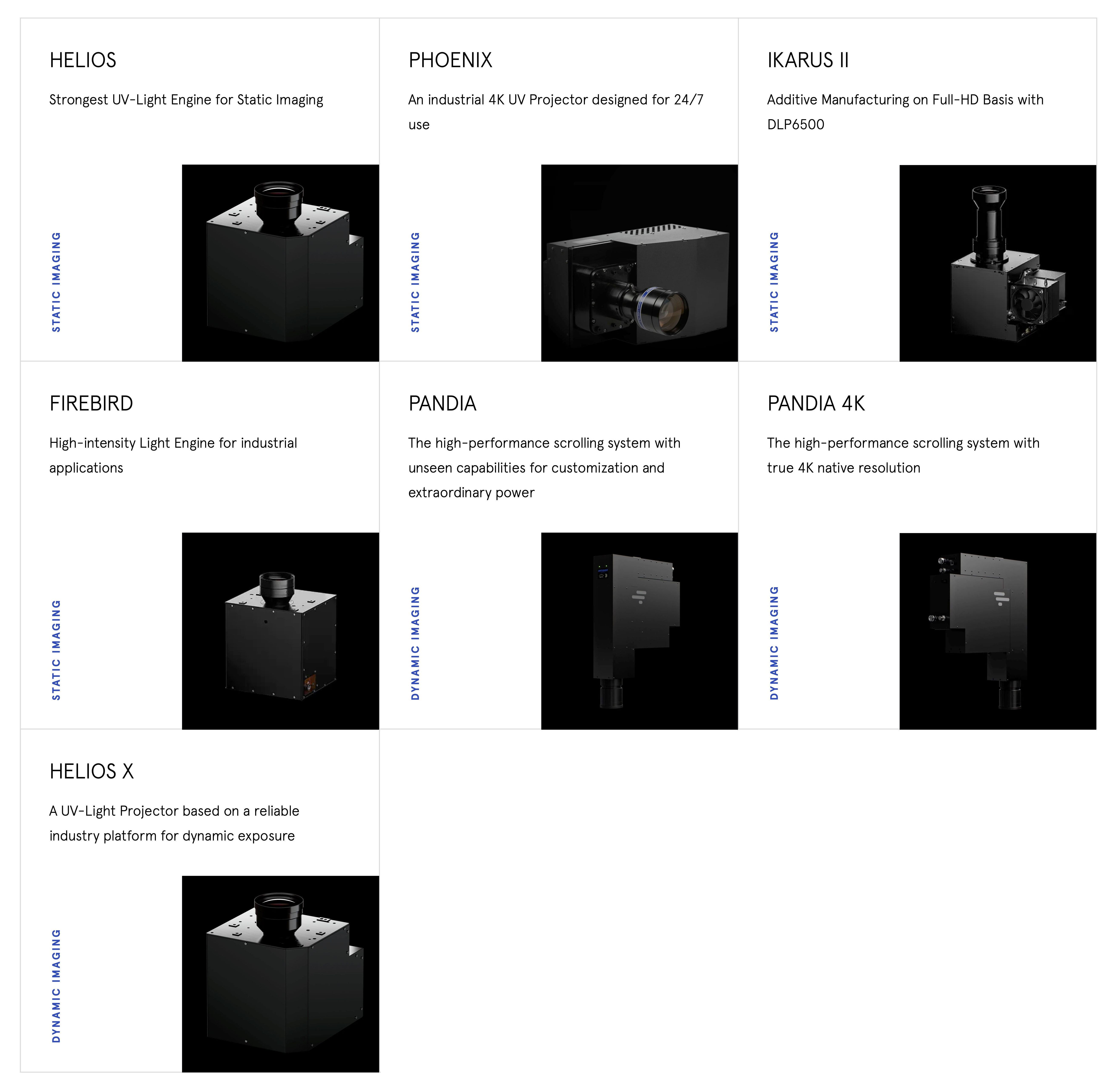
In-Vision’s Light Engines are designed for industrial applications fulfilling the requirements of a 24/7 reliable UV projector with small pixel sizes.
What if we could simply regrow an organ, using the patient’s own cells?
Bioprinting provides exciting avenues to solve these challenges. Inspired by additive manufacturing technologies, researchers and technology pioneers have set out to print (human) tissues and organs. A biocompatible polymer is printed to provide the support matrix, into which living cells are seeded to grow into an organ. Via multi-material printing, the complex geometries of different cell types forming a functional organ are replicated. Seeded with patient-derived cells, such an organ would not be attacked by the immune system anymore. For any drug development, animal testing remains an integral step, despite limited transferability of the obtained results – mice are simply not identical to humans. Ethical and moral concerns dictate that we reduce animal experimentation to a minimum. Even in human trials, rare side effects are often overseen.
What if we could estimate patient-specific side effects of a treatment before administering it?
Using microfluidics technology, researchers have been trying to reduce the structural complexity of organs, while still replicating the functionality. In a “organ-on-a-chip”, liquids are running through tiny fluidic channels, separated by membranes. The shape of the channels, as well as the type of cells seeded onto the membranes, determine how the liquids are mixed or separated, replicating the function of kidneys, lungs, skin, or the gut. In a organ-on-a-chip populated with the patient’s cells, it will become to test for patient-specific effects of a treatment – completely in vitro, without the need for testing in living animals or humans. With 3D printing, the design flexibility for the structure of the channels greatly improved over classical lithography masks.
The challenges posed by biological system are enormous, and the safety and reliability requirements of any medical application are strict.
Related Articles
- "When will the first organ come out of the printer?" Interview with Bioprinting pioneer Dr. Y. Shrike Zhang from Harvard University
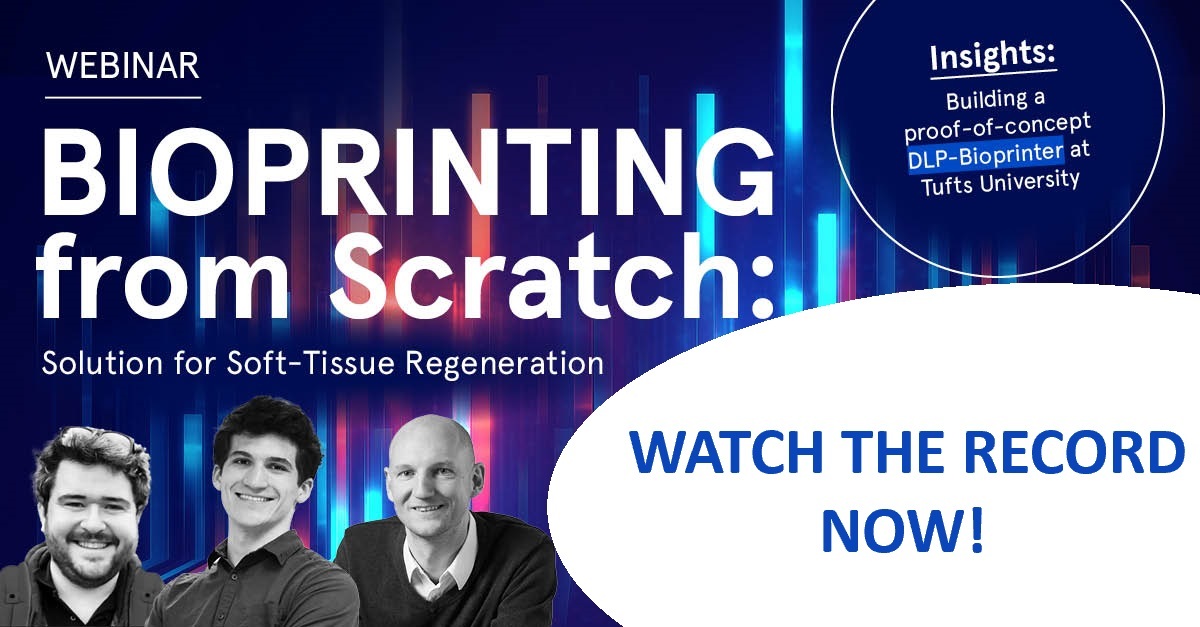
- Advanced Soft Tissue Regeneration - Process of building a proof-of-concept bioprinter with In-Vision's Ikarus Light Engine
- "There is still so much to explore" - Wiebke Jahr, project manager at In-Vision, shares her personal experiences and opinion about optics and life sciences
- Bioprinting explained
- Biocompatibility explained


